

Musculoskeletal disorders affect millions of people worldwide, ranging from acute sports injuries and tendon tears to chronic conditions such as arthritis and repetitive strain injuries. Accurate imaging plays a critical role in diagnosing these conditions, monitoring recovery, and guiding treatment decisions. As healthcare providers continue to seek faster, safer, and more accessible diagnostic tools, musculoskeletal ultrasound imaging has become an increasingly important part of modern musculoskeletal care.
In this article, we’ll explore how musculoskeletal ultrasound imaging works, its most common clinical applications, the factors driving its growing adoption, and the latest developments shaping its future.

What Is Musculoskeletal Ultrasound Imaging?
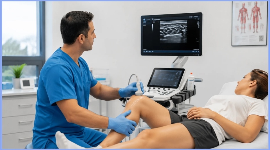
Musculoskeletal ultrasound imaging (MSK ultrasound) is a non-invasive diagnostic imaging technique that uses high-frequency sound waves to generate real-time images of the body’s musculoskeletal structure. It enables healthcare providers to evaluate soft tissues without exposing patients to ionizing radiation.
Unlike imaging modalities that produce only static images, musculoskeletal ultrasound imaging allows clinicians to observe tissues in real time as a patient performs specific movements. This capability makes it particularly useful for evaluating conditions that become apparent only during motion. In addition to its diagnostic value, ultrasound is frequently used to guide minimally invasive procedures, including joint aspirations, corticosteroid injections, and nerve blocks, improving procedural accuracy while minimizing the risk of injury to surrounding structures. Its combination of real-time visualization, accessibility, and excellent soft-tissue resolution has made musculoskeletal ultrasound imaging an essential component of modern orthopedic, sports medicine, rheumatology, and rehabilitation practices.
How Does Musculoskeletal Ultrasound Imaging Work?
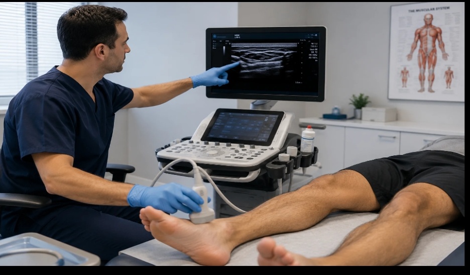
Musculoskeletal ultrasound imaging uses a handheld transducer to emit high-frequency sound waves into the body. As these waves pass through muscles, tendons, ligaments, nerves, and other soft tissues, they are reflected back at different intensities depending on the characteristics of each structure.
The ultrasound machine processes these returning echoes and instantly converts them into detailed, real-time images displayed on a monitor. Because ultrasound uses sound waves rather than ionizing radiation, it is considered a safe imaging modality for repeated examinations and ongoing patient monitoring.
As mentioned earlier, one of the greatest strengths of musculoskeletal ultrasound imaging is its ability to perform dynamic assessments. This means that clinicians can evaluate muscles, tendons, ligaments, and joints as patients move the affected area, making it easier to identify conditions that may not be visible on static imaging. High-frequency linear transducers such as the Sonosite HFL38x are typically preferred because they provide excellent resolution for superficial structures, helping clinicians visualize subtle tissue abnormalities with greater clarity.
Common Applications of Musculoskeletal Ultrasound Imaging
Musculoskeletal ultrasound imaging is used across a wide range of medical specialties to evaluate soft tissue injuries, monitor chronic musculoskeletal conditions, and guide minimally invasive procedures. Some of its most common applications include:
- Tendon Injuries: Evaluates tendon tears, tendinopathy, and inflammation in the rotator cuff, Achilles tendon, and patellar tendon.
- Ligament Injuries: Helps identify sprains, partial tears, and complete ligament ruptures while allowing dynamic assessment of joint stability.
- Muscle Disorders: Detects muscle strains, tears, hematomas, and other soft tissue abnormalities, particularly in sports-related injuries.
- Joint Disorders: Assesses joint effusions, synovitis, bursitis, and other inflammatory conditions affecting the shoulder, knee, hip, wrist, and ankle.
- Peripheral Nerve Evaluation: Visualizes nerve compression and entrapment syndromes, including conditions such as carpal tunnel syndrome and ulnar neuropathy.
- Sports Medicine: Supports the diagnosis and monitoring of acute and chronic athletic injuries, enabling clinicians to evaluate tissue healing throughout rehabilitation.
- Rheumatology: Assists in detecting inflammatory changes in joints, tendons, and surrounding soft tissues associated with diseases such as rheumatoid arthritis.
- Ultrasound-Guided Procedures: Provides real-time guidance for joint aspirations, corticosteroid injections, platelet-rich plasma (PRP) therapy, and peripheral nerve blocks, improving procedural accuracy and patient safety.
These diverse applications have made musculoskeletal ultrasound imaging an essential diagnostic and interventional tool in orthopedic clinics, sports medicine centers, rehabilitation facilities, rheumatology practices, and pain management clinics worldwide.
Why Musculoskeletal Ultrasound Imaging Has Become More Popular
The increasing adoption of musculoskeletal ultrasound imaging reflects broader changes in modern healthcare. As clinicians place greater emphasis on early diagnosis, minimally invasive treatments, and efficient patient care, ultrasound has become an increasingly practical imaging solution. It enables healthcare providers to examine patients during a single appointment, helping reduce delays between assessment, diagnosis, and treatment while improving overall clinical workflow.
Another key factor behind its growing popularity is the continuous advancement of ultrasound technology. Modern systems provide higher image quality, greater portability, and AI-assisted features that enable faster, more consistent examinations. At the same time, growing clinician experience and expanded training in musculoskeletal ultrasound have increased confidence in its use across specialties such as orthopedics, sports medicine, rheumatology, and rehabilitation. Together, these developments have established musculoskeletal ultrasound imaging as an integral part of contemporary musculoskeletal care.
PhD lecturer enrolled at KMTC campus, studying Diploma in Clinical Medicine
Risks and Benefits of Musculoskeletal Ultrasound Imaging
Like any medical imaging modality, musculoskeletal ultrasound imaging has both advantages and limitations, as shown in the table below.
| Benefits | Risks |
| No ionizing radiation | Operator-dependent |
| Real-time imaging | Bone limitations |
| Excellent soft tissue visualization | Smaller field of view |
| Portable and accessible | Some conditions require additional imaging |
The Continued Growth of Musculoskeletal Ultrasound Imaging in 2026 and Beyond
Musculoskeletal ultrasound imaging is expected to play an even greater role in healthcare as technology continues to advance and clinical expertise expands. Improvements in image quality, high-frequency transducers, artificial intelligence, and portable ultrasound systems are making examinations faster, more accurate, and more accessible across a wide range of healthcare settings. At the same time, growing demand for point-of-care imaging and minimally invasive procedures is encouraging more hospitals, outpatient clinics, and sports medicine centers to incorporate ultrasound into routine musculoskeletal assessments.
Looking ahead, continued investment in ultrasound technology is likely to further enhance diagnostic capabilities and clinical workflows. Better software integration, cloud-based data management, and AI-assisted image analysis are expected to improve efficiency while supporting more consistent clinical decision-making. As these innovations become increasingly available, musculoskeletal ultrasound imaging will continue to strengthen its position as a valuable diagnostic tool for evaluating soft tissue conditions and guiding patient care across multiple medical specialties.
Conclusion
Musculoskeletal ultrasound imaging has become an essential tool for evaluating a wide range of musculoskeletal conditions, offering clinicians a safe, real-time, and highly effective method for examining soft tissues and guiding minimally invasive procedures. Continuous advancements in ultrasound technology, including higher-resolution imaging, high-frequency transducers, and AI-powered features, have expanded its clinical capabilities while improving diagnostic confidence and workflow efficiency.
As healthcare providers continue to prioritize earlier diagnosis, personalized treatment, and point-of-care delivery, the role of musculoskeletal ultrasound imaging is expected to grow even further. Choosing the right imaging equipment and transducer is an important part of achieving optimal clinical outcomes, ensuring healthcare professionals can deliver accurate diagnoses and high-quality patient care across a broad range of musculoskeletal applications.